Bitemporal hemianopsia: diagnosis and treatments

The editorial staff of Emianopsia is pleased to propose the interview to Dr Borghetti: graduated in Medicine and Surgery at the University of Pisa, specializing in Neurology. Over the years he has gained extensive experience in the field of neurological diagnostics, regarding electrophysiological and magnetic resonance studies. We thank him for making his experience available to readers to clarify symptomatology that differs in: bitemporal hemianopsia, binasal, homonymous and altitude.
Can you tell us what is the experience gained with patients with bitemporal hemianopsia, homonymous, altitudinal, binasal? What is your most common clinical picture?
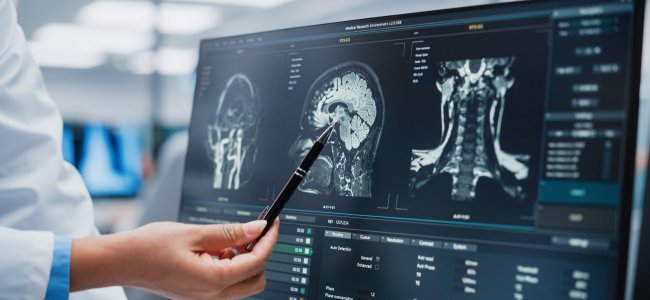
“Over the years, I have had the opportunity to deal with hemianopsia both on the strictly clinical, ( therefore neurological and neuroophthalmological side ), and on the instrumental one. Modern neuroimaging techniques – such as magnetic resonance imaging, with which I have had a long experience – are characterized by high levels of sensitivity, essential to reach the correct and definitive diagnosis of said disorder.
Among the most frequent forms, there are undoubtedly those related to outcomes of a vascular (haemorrhagic or ischemic), neoplastic (even histologically benign) and post-traumatic nature. There is, however, the possibility that hemianopsia is secondary or relatively rarer situations, such as infectious events or even recognize a post-surgical (iatrogenic) genesis.
It should also be taken into account that, many of these patients have comorbidities linked to the possible extension of the lesion to other anatomic-functional brain sections. It is therefore not uncommon to observe the presence of other neurological disorders, such as changes in walking, sensitivity, coordination, speech and so on“.
What is the patient’s awareness of their visual impairment? What are their demands and needs?
“This may sound peculiar, but not all people with hemianopsia have a clear perception of their deficit. In some cases, even, this emerges only during an occasional check, perhaps carried out for other reasons. Most people, however, soon realize that something is wrong, reporting a vague disturbance of vision that will lead them, later, to start a complete diagnostic process.
Still, others report with disarming clarity the characteristics of their deficit (“I cannot see half of the world“) and are even able to draw a clear line between what they see and what they fail to perceive. Many people realize that they can no longer do simple things like reading or watching a movie, as the deficit makes it difficult to properly explore the environment.
Others still bump into furniture and objects, especially in unfamiliar environments, or even realize they can no longer drive, write, or simply cross the road with serenity. The needs and requests of the patient, therefore, are focused on the recovery of their autonomy, also and above all for small daily activities. “
Hemianopsia, being very peculiar as symptomatology, hides pitfalls during the diagnosis process (it may be difficult to make a distinction between this and neglect). Can you explain to us how the diagnosis is carried out and what are the signs that assimilate the visual deficit to the hemianopsia?
“In some cases, the differential diagnosis between neglect (hemi inattention) and hemianopsia can be difficult. It becomes therefore essential not to make hasty decisions and carefully evaluate the characteristics and data of the patient and about the disorder.
The first element to be considered is that of the distribution of brain injury, which must be in line with the suspected deficit. In general, moreover, hemianopsia is (but not necessarily) an isolated disorder, while neglect is more frequently associated with other deficits (e.g. auditory, tactile or motor). This will depend, to a large extent, on the causes and actual distribution of the brain injury.
In neglect, by definition, there is inattention of the contralateral hemisphere, which occurs regardless of the direction. In hemianopsia, on the other hand, the deficit strictly follows the direction of the eyes: some objects can be observed simply by moving the eyes. In this case, moreover, the subject does not usually have difficulty in looking for a prolonged amount of time at the same thing or at the centre of their visual field, which is instead much more difficult in neglect “
Regarding recovery from hemianopsia, multiple approaches have been developed (for example, the neuro-rehabilitative approach, which exploits optical aids such as prismatic lenses). Could you tell us the benefits and issues that characterize them?
“Rehabilitation approaches in hemianopsia can be divided into three main groups. To begin with, we have training aimed at ameliorating the ocular movements of exploration, that is, the behaviour that leads us to explore all the space around us by moving the direction of the gaze. If the field of view is lacking (as in hemianopsia) it is however possible to make a satisfactory overview simply by reorganizing the way we explore the said environment.
Prisms, on the other hand, are used to expand the field of view: this is feasible only if the prismatic image can be put within the patient’s residual view field. Unfortunately, prismatic lenses represent a real challenge (both in terms of realization and tolerance) and are not free from problems, among which the risk of “split” vision, which however tends to reduce over time.
The neuro-rehabilitative approach, finally, exploits the mechanisms of neural plasticity and integration of multisensory elements, such as visual and auditory stimuli. This results in a progressive reinforcement of compensatory functions (such as the implementation of adequate eye care strategies).”
We thank Dr Borghetti for giving us a neurological point of view on such peculiar symptomatology as homonymous, altitudinal, binasal, and bitemporal hemianopsia. Continue reading the interview with Dr Borgetti: Cognitive deterioration: the rehabilitation path.

You are free to reproduce this article but you must cite: emianopsia.com, title and link.
You may not use the material for commercial purposes or modify the article to create derivative works.
Read the full Creative Commons license terms at this page.


