Congenital Hemianopsia: consequences for daily life

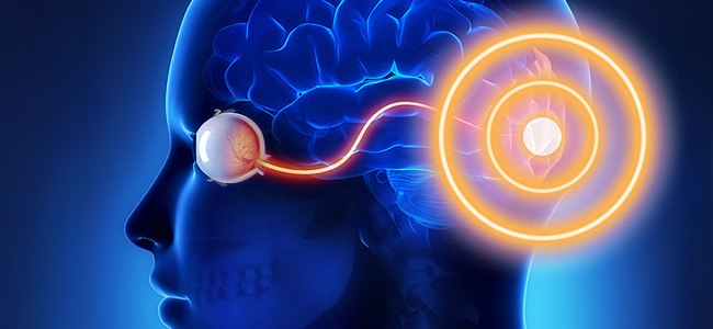
Congenital hemianopsia represents a vision condition characterized by the reduction or complete absence of perception of half of the visual field.
Hemianopsia is a visual disorder characterized by the reduction or complete absence of the perception of half of the visual field. Visual field loss can be lateral hemianopsia (right or left), altitudinal hemianopsia (above or below), or horizontal hemianopsia (either right or left).
In heteronymous hemianopsia, there is a loss of the outer or inner two halves of the visual field. This is a disease called bitemporal heteronymous hemianopsia.
Hemianopsia can be further divided into two categories: heteronymous hemianopsia and homonymous hemianopsia, based on the location of visual field loss.
It causes the loss of the two outer halves of the visual field. This can be caused by a lesion or compression of the optic pathways in the region of the optic chiasm. This type of hemianopsia may be associated with pathologies such as pituitary adenomas or tumors affecting the optic chiasma.
In basal heteronymous hemianopsia, however, there is a loss of both inner halves of the visual field. This type of hemianopsia is caused by a lesion or compression of the optic pathways.
This lesion or compression occurs in the region of the optic tract. Visual information fails to reach the optic chiasm. Pathologies such as aneurysms or tumors in this region may be responsible for basal hemianopsia.
In basal hemianopsia, there is a loss of either the left or right halves of the visual field. This condition can be caused by a lesion or compression. The affected areas are the optic pathways or cortical visual areas on either the left or right side of the brain. For example, a stroke or brain injury involving the left-sided occipital cortex can result in left homonymous hemianopsia.
The distinction between heteronymous hemianopsia and homonymous hemianopsia is important as it can provide clues to the site and cause of optic pathway injury or compression. Accurate diagnosis of the type of hemianopsia is critical to the selection of appropriate treatment and management of the patient’s visual condition.
Causes
Congenital hemianopsia is a rare visual condition that occurs from birth, causing partial or complete loss of the visual field in one or both eyes. Its causes can vary and include congenital defects of the optic pathways or visual cortex. Despite its rarity, the medical literature offers data and research that delve into the possible causes of congenital hemianopsia.
A study of infants revealed that about 1-3% of congenital hemianopsia cases can be attributed to congenital defects of the optic pathways. These defects may involve failure to form or abnormal development of the optic pathways, interfering with the transmission of the visual signal.
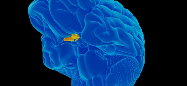
An analysis of epidemiological data suggested that congenital defects of the visual cortex may cause congenital hemianopsia. These defects are responsible for less than 1% of cases. These abnormalities in the formation of the visual cortex may affect the proper processing of visual information, leading to visual field loss.
However, it is important to note that statistics on congenital hemianopsia may vary depending on the studies and sources consulted. It is crucial to consider that specific causes may differ from case to case, and the etiology of congenital hemianopsia requires an accurate and thorough diagnosis. Therefore, a personalized approach to the management of this congenital visual condition is essential to ensure appropriate care for affected patients.
Challenges in daily life
Congenital hemianopsia, a condition characterized by visual field limitation, can affect several aspects of daily life. People with congenital hemianopsia may experience difficulties in spatial orientation and perception of surrounding space, making walking and obstacle avoidance complex. In addition, impaired ability to recognize objects or people in peripheral vision may impair road safety while driving. Activities such as reading, watching movies, or attending social events may require additional efforts to compensate for visual field loss.
To cope with congenital hemianopsia, several strategies affected persons can adopt. The use of visual landmarks, adequate lighting, and the implementation of routines that promote greater awareness of one’s surroundings can improve spatial orientation. Visual aids, such as blind spot mirrors or prismatic glasses, can expand the useful visual field and facilitate the compensation of blind spots.
Visual rehabilitation plays an important role in supporting people with congenital hemianopsia. Rehabilitation programs may include specific exercises aimed at improving spatial awareness, orientation, and blind spot compensation. Visual rehabilitation specialists provide individualized support and teach specific strategies for coping with the daily challenges associated with congenital hemianopsia.
Congenital hemianopsia can have a significant impact on the emotional and social aspects of those affected. Visual difficulties can cause frustration, anxiety, or social isolation. Therefore, it is important to seek psychological support and connect with support groups, where people can share experiences and strategies with individuals experiencing the same condition. Family and friends’ support plays a key role in providing emotional and practical support in coping with congenital hemianopsia.
In conclusion
In conclusion, congenital hemianopsia poses a significant challenge for those affected, affecting various aspects of their daily lives. People with this condition can manage and overcome the difficulties that come with it. This is possible with the help of proper compensatory strategies and support.
The use of visual landmarks, visual aids such as blind spot mirrors and prismatic glasses, along with targeted visual rehabilitation programs, can help improve spatial orientation and compensate for blind spots.
In addition, psychological support, interaction with support groups, and family and social support are crucial in coping with the emotional and social impact of congenital hemianopsia.
People with congenital hemianopsia need adequate access to resources. A multidisciplinary approach involving visual rehabilitation specialists, mental health professionals, and social support is essential. This will enable them to find effective ways to cope with daily challenges. Thus, they can lead fulfilling lives despite their limiting visual condition.
References:
- Mandelstam, S. A., Maixner, W. J., Grigg, J. R., & Graham, S. L. (2013). Congenital Visual Pathway Abnormalities: A Review. Clinical & Experimental Ophthalmology, 41(4), 397-407.
- Apkarian, P., Reits, D., & Spekreijse, H. (2000). Visual field defects in children: a review. Documenta Ophthalmologica, 100(2-3), 191-207.
- Baker, D. H. (2015). Congenital Hemianopia. In B. Simons (Ed.), The Oxford Handbook of Visual Cognition (pp. 697-716). Oxford University Press.
- Chokron, S., & Perez, C. (2016). Rehabilitation of homonymous hemianopia: insight into blindsight. Frontiers in Integrative Neuroscience, 10, 31.
- Fletcher, D. C., Schuchard, R. A., & Watson, G. (2016). Relative locations of macular scotomas near the PRL: Effect on low vision reading. Journal of Vision, 16(8), 8.
- Geruschat, D. R., & Turano, K. (2018). Living with vision loss: The impact on the individual and society. In A. Arditi & E. L. B. Best (Eds.),
- The Ophthalmic Assistant: A Text for Allied and Associated Ophthalmic Personnel (pp. 737-747). Elsevier.

You are free to reproduce this article but you must cite: emianopsia.com, title and link.
You may not use the material for commercial purposes or modify the article to create derivative works.
Read the full Creative Commons license terms at this page.


